
紹介患者さんの治療と経過観察。
主訴は、
左上奥歯, 今は痛くないが紹介された時には痛みがひどく, ものが噛めなかった…
である。
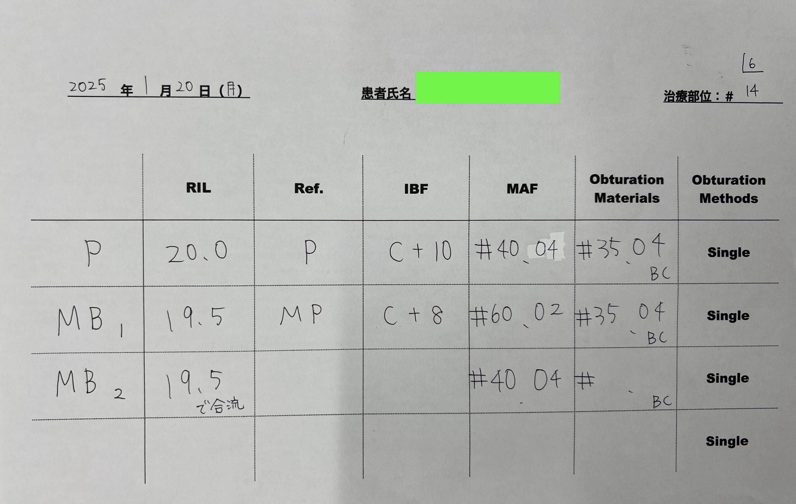
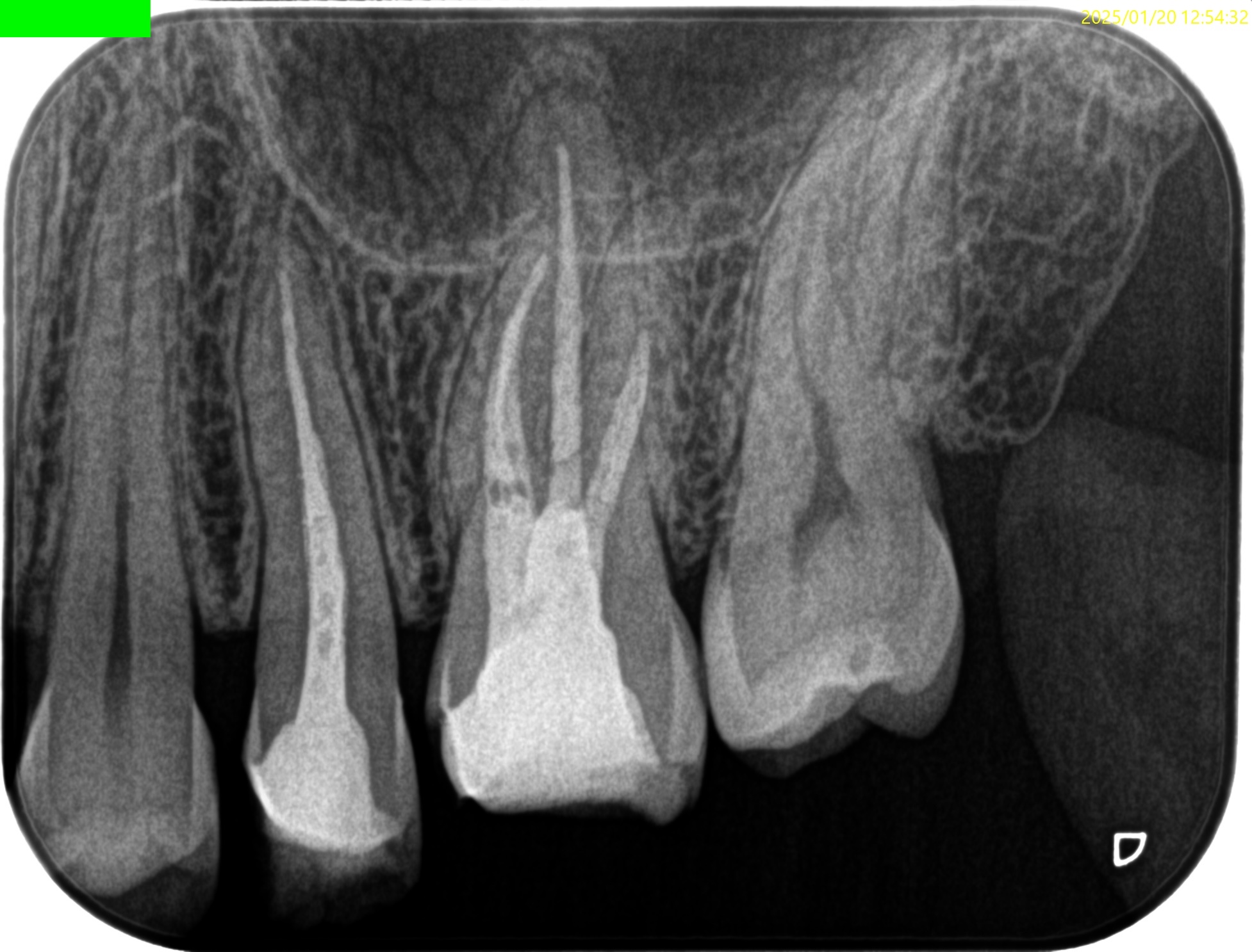
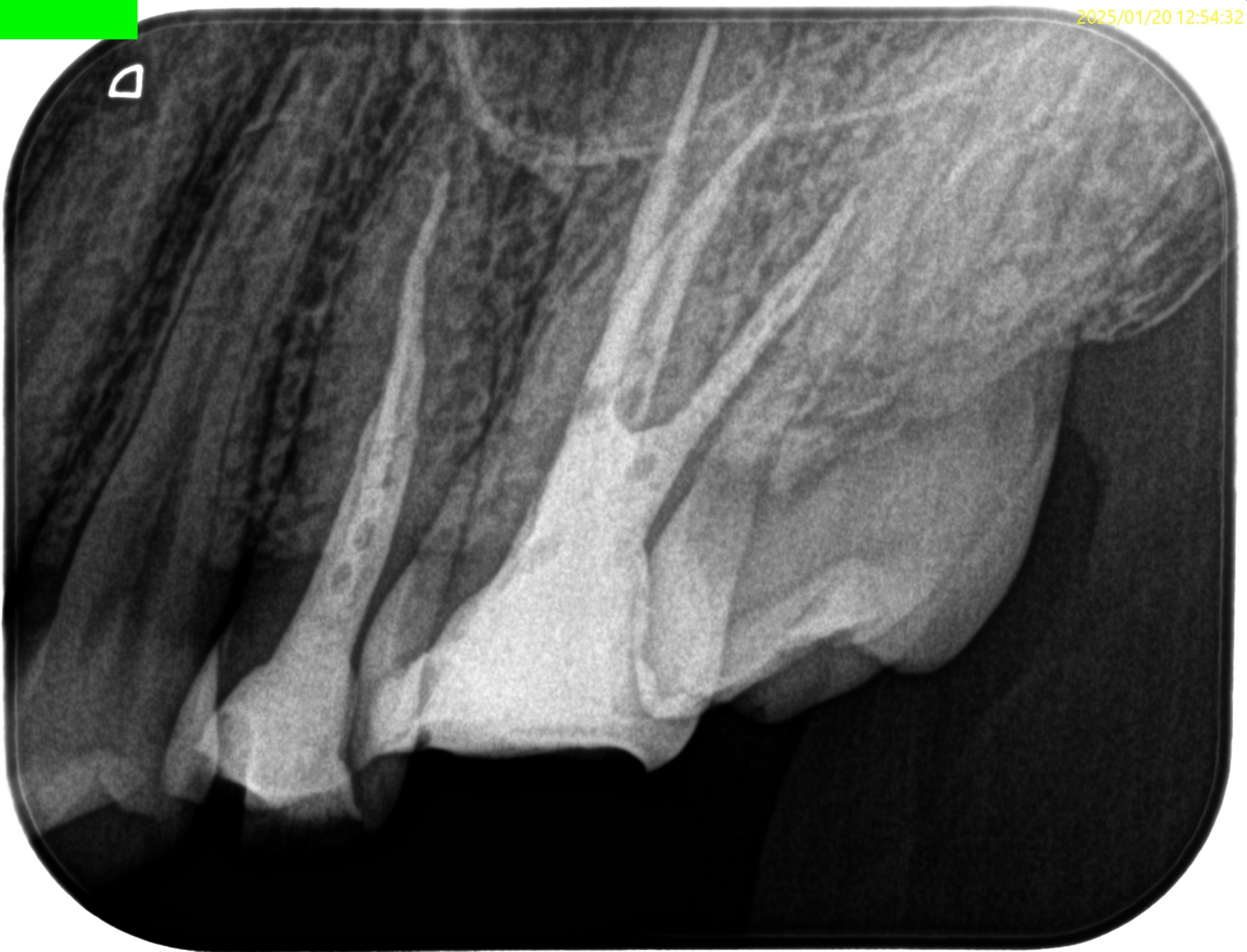
Pre-op Endo test(2025.1.20)
臨床症状はないが、
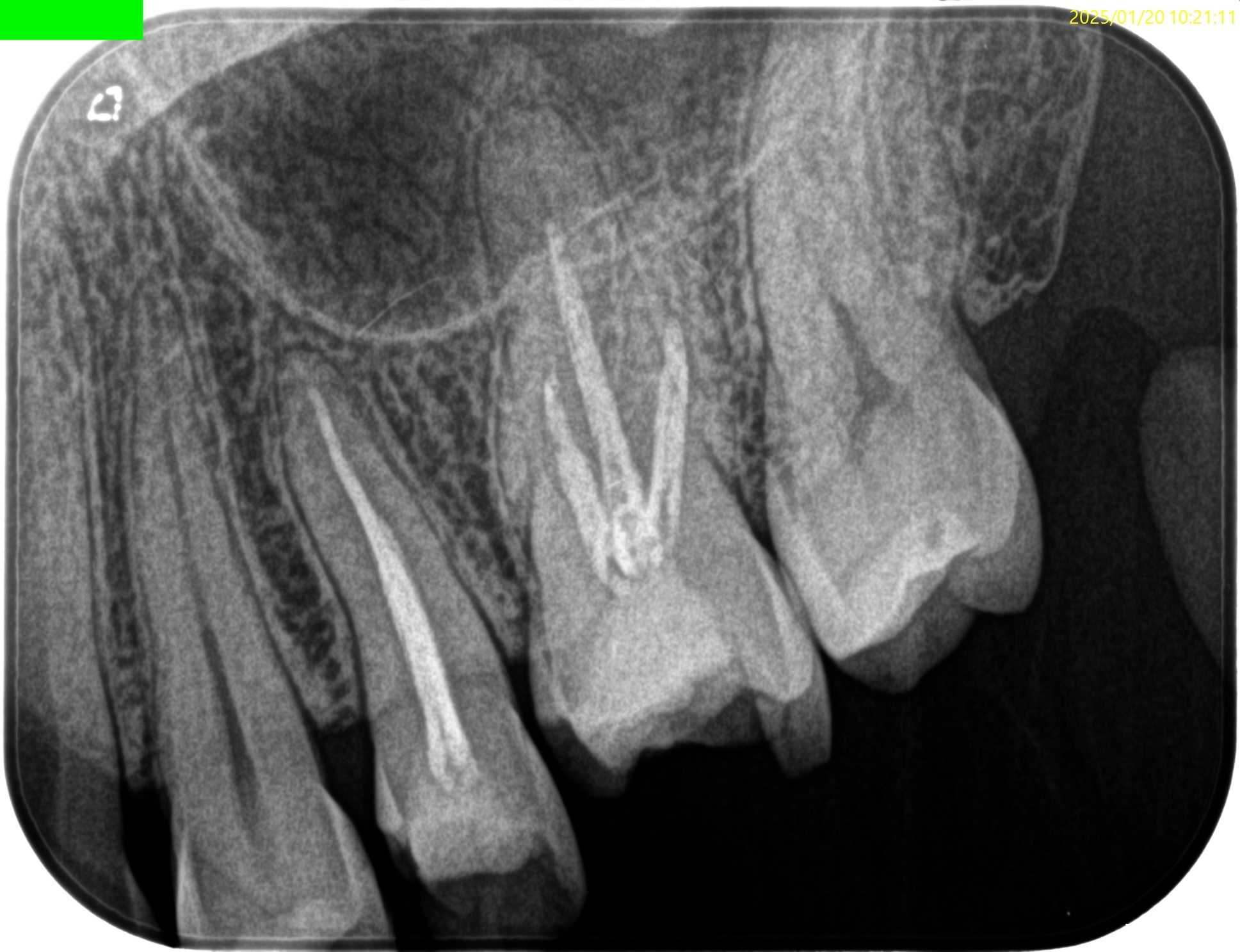
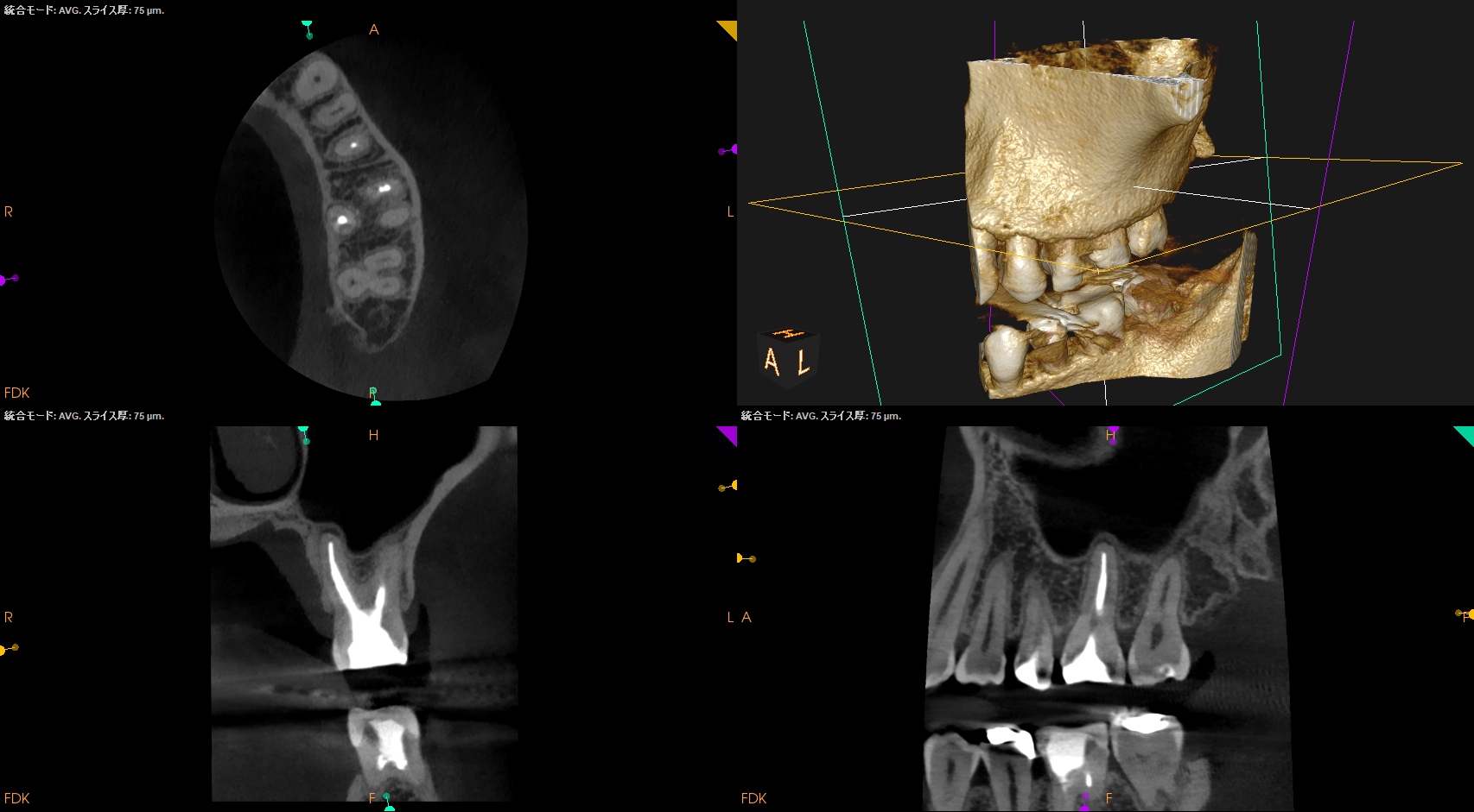
#13
#13に根尖病変はない。
補綴には築造のみ必要だろう。
#14
MB1

MB2
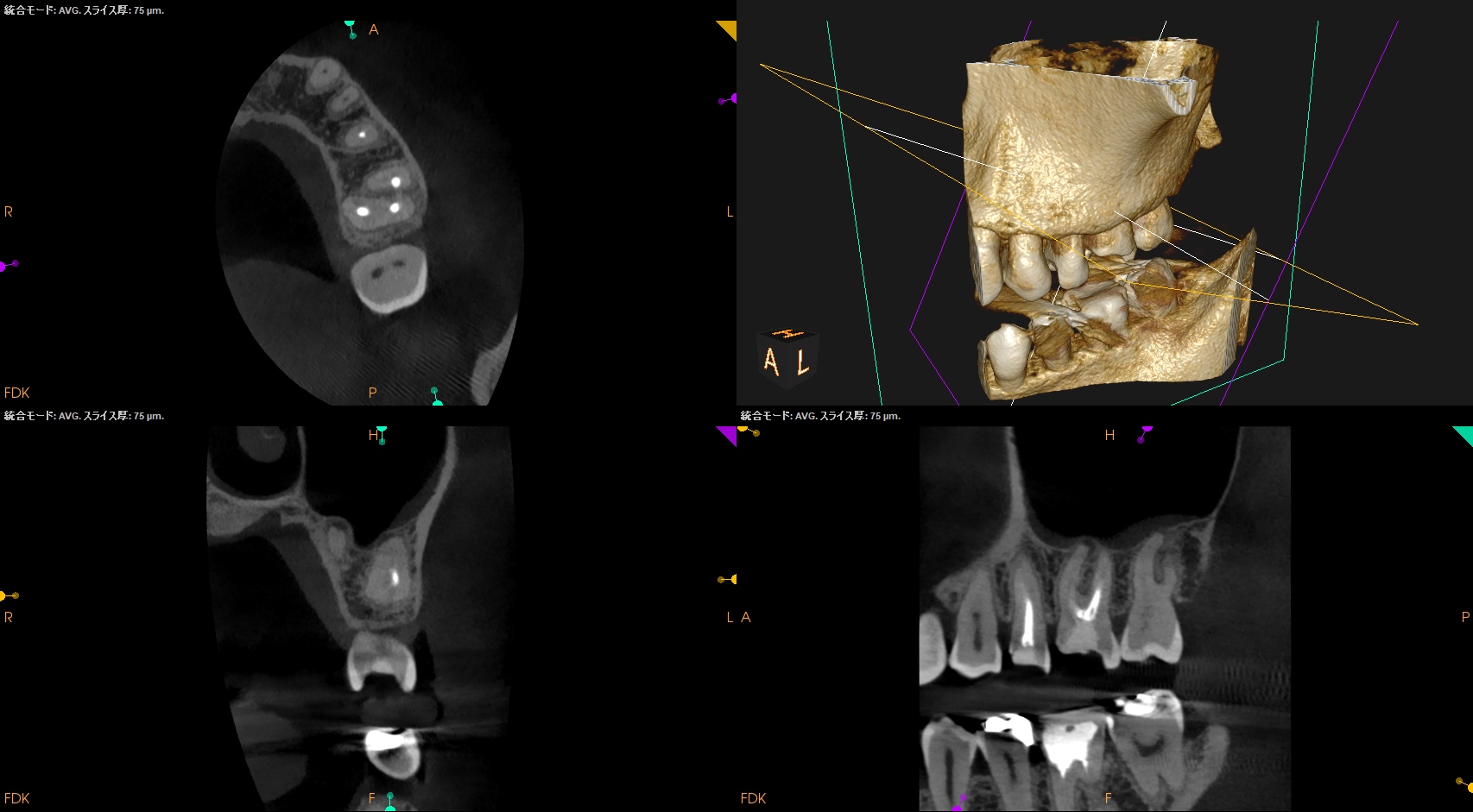
MB2は未着手でその根尖に病変がある。ここは発見と根管形成が必要だ。
DB
DBには病変がない。
根管充填は疎だが基本, 触れる必要もなさそうだ。Selectiveに再治療を行なっていくと決めた。
そんなことをして大丈夫なのか?
大丈夫かはCaseを見て判断するしかない。
Sinus tractがある石灰化が亢進した歯牙の再根管治療(Selective Root Canal Re-treatment)〜#30 Re-RCT 1回法 8M recall
P

Pにも病変が見られる。
そしてほぼ形成していない。
以上のことから、
再根管形成が臨床的に必要なのはMB2とPであるということがわかる。
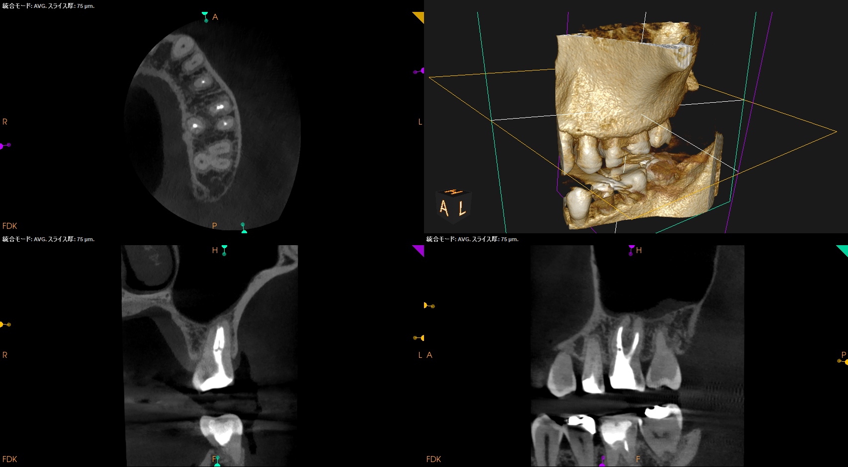
Pre-op Endo Diagnosis(2025.1.20)
Pulp Dx: Previously initiated therapy
Periapical Dx: Asymptomatic apical periodontitis
Recommended Tx: Re-RCT
では、MB2はどこにあるのだろうか?
上顎第1/第2大臼歯のMB2に関しては以前の記事でも触れている。
☆How to find MB2?
1. Locate the Position: The MB2 is generally found 1–3 mm palatal to the main MB1 canal, not directly between the MB1 and Palatal canals, but often on a line connecting them
2. Remove the Dentin Shelf: A significant portion of the MB2 orifice is covered by a “white line” or a shelf of dentin. Use ultrasonic tips or a small, long-necked, round bur to trough 1–2 mm deep into the Mesial-Palatal developmental groove.
3. Use Magnification & Lighting: A dental microscope or high-powered loupes are essential, as the MB2 is frequently too small or hidden to see with the naked eye.
4. “Troughing” Technique: Use an ultrasonic tip (e.g., Munce Discovery) to trough along the pulpal floor between the MB1 and the palatal orifice.
5. Identify Bleeding Points: A “blood trace” or small pinpoint bleed on the pulpal floor, especially after removing debris, often indicates the location of the MB2.
6. Use Dyes: Methylene blue or another dye can help highlight the pulpal floor map.
7. Initial Exploration: Once located, use a small, hand-held file (e.g., #06 or #08) or a micro-opener with ultrasound to navigate the first few millimeters.
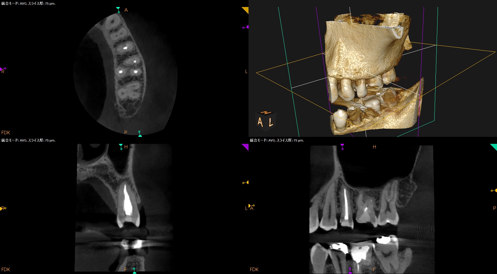
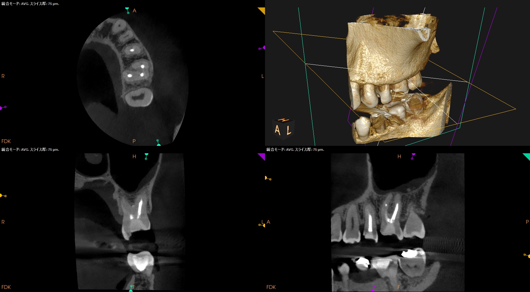
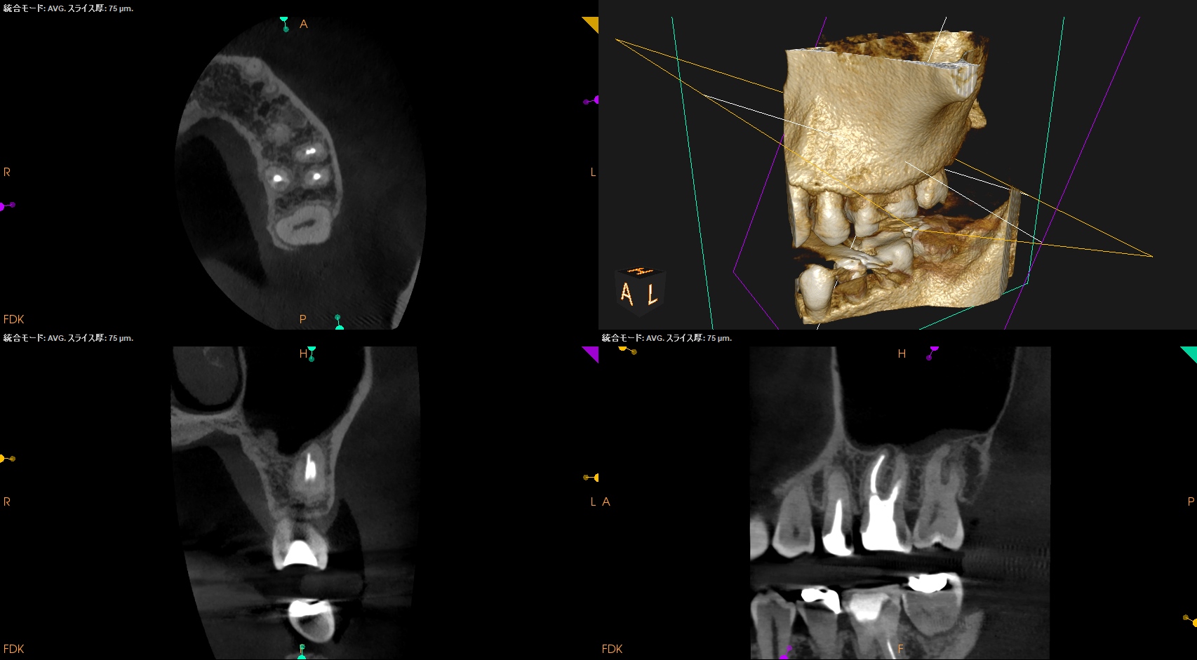
CBCTを参考にすると、MB2のすぐ後ろに根管口と思しき点が見える。
そこがMB2の根管口だろう。
が、それを見つけること、見つかること
と
穿通して根管形成, 根管充填を成功させることができるか?
は
別問題である。
ここを誤解してはいけない。
ということで、同日に治療へ移行した。
☆この後、治療動画が出てきます。不快感を感じる方は視聴をSkipしてください。
#14 Re-RCT(2025.1.25)
MB2はMB1を形成してから探したほうが効率がいい。
その理由はその多くは合流している可能性が高いからだ。
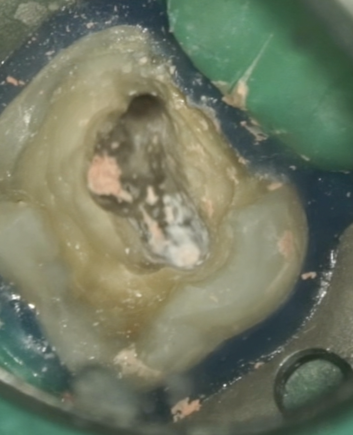
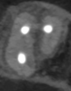
この絵のどこにMB2があるか?あなたは予想がつくだろうか?
一般的にはその法則は以下だ。
MB2 Detection Strategy
①Using CBCT imaging, the ratio between the distance from the MB1 to the P (MB-P) and the distance from the DB to P (DB-P) may help predict the presence of MB2. When MB-P / DB-P> 1.26, the likelihood of an MB2 canal is very high.
②First, locate the MB and P canal orifices and draw a line connecting them. Then draw a perpendicular line from the DB canal. The MB2 canal is likely located at the intersection of these two lines.
③In most cases, the line connecting MB2 and MB is parallel to the mesial marginal ridge.
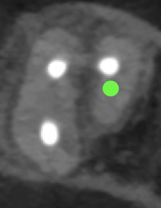
以上の情報を上記画像に当てはめると、MB2の当該部位は以下だろう。
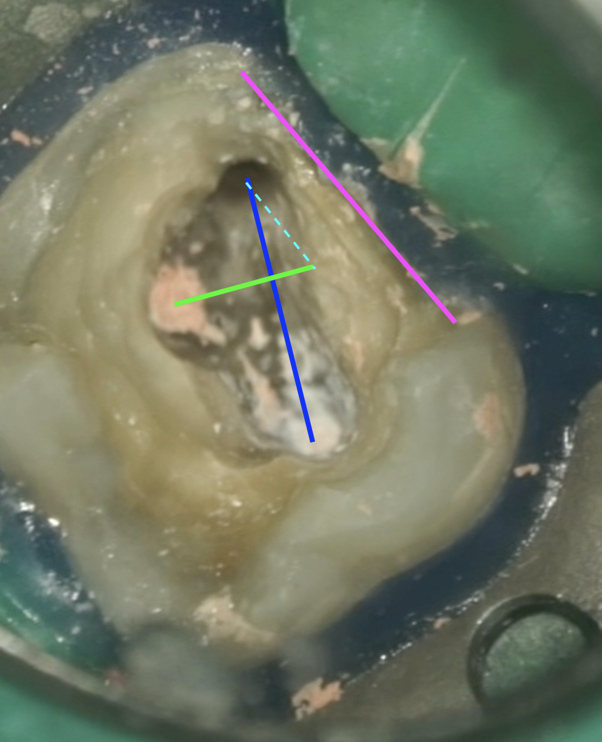
上記絵を参考にMB2を探索してみた。
見事MB2は見つかりSXで形成を行うこともできた。
MB2の多くはMB1と合流することが知られている。
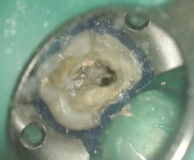
以下が作業内容である。
術後にPA, CBCTを撮影した。
MB1
MB2
P
次回は1年後である。
またその模様をお伝えしたい。